At Ollins Orthodontics, digital imaging and x-rays are integral aspects of our practice, offering unparalleled accessibility and precision in patient care. Today, we’ll delve deeper into these essential instruments and answer this question: How are x-rays and imaging used to track progress in orthodontics?
About X-rays & Imaging
There are multiple methods we can utilize in imaging for our patients based on their condition.
- Intraoral X-rays: This method is the most prevalent in dental imaging, involving the placement of a small sensor or film inside the patient’s mouth to capture detailed images of individual teeth, roots, and surrounding bones. These x-rays play a vital role in assessing bone levels, detecting cavities, and evaluating the health of tooth roots.
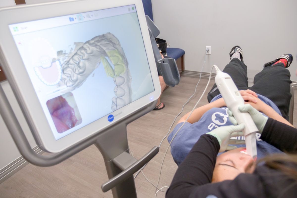
- Digital Imaging: Utilizing digital sensors and intraoral cameras, this technique captures high-resolution images of the teeth and gums. These images are promptly available for viewing on a computer screen, enabling dentists to visualize dental conditions, educate patients, and plan treatment approaches more effectively.
- Panoramic X-rays: These provide a comprehensive perspective of the entire mouth, encompassing all teeth, upper and lower jaws, and adjacent structures. They are invaluable for assessing overall dental health, identifying impacted teeth, and formulating treatment plans for orthodontic care or oral surgery.
Imaging in Action
Now that you know what some of these processes look like, we can discuss how they’re applied in our practice.
- Initial Diagnosis and Treatment Planning: This imaging is pivotal during the initial stages of diagnosis and treatment planning. It provides detailed insights into tooth alignment, jaw relationships, the presence of impacted teeth, and any underlying skeletal abnormalities. During your first exam, we’ll usually use digital imaging.
- Monitoring Tooth Movement: X-rays and imaging are utilized to monitor the progression of tooth movement throughout orthodontic treatment. Serial periapical or panoramic radiographs are taken at regular intervals to assess tooth positions and track their movement over time.
- Evaluation of Root Resorption: We can evaluate the degree of root resorption, which refers to the shortening of tooth roots that may occur during orthodontic treatment. By periodically assessing root length and morphology, orthodontists can identify signs of excessive root resorption and adjust treatment as necessary to prevent further damage.
- Assessment of Treatment Stability: Following treatment completion, both x-rays and imaging are employed to evaluate the stability of treatment outcomes. By comparing post-treatment images with baseline images taken before treatment, our doctors can discern any changes and decide if additional interventions are necessary to uphold results.
- Identification of Orthodontic Emergencies: X-rays are instrumental in identifying and diagnosing orthodontic emergencies, such as loose brackets, broken wires, or dislodged teeth. By visualizing the underlying tooth and jaw structures, x-rays aid in determining the appropriate steps to promptly and effectively address these issues.
- Communication with Patients and Referring Providers: Imaging offers visual depictions of a patient’s dental and skeletal anatomy, facilitating communication with patients and referring providers regarding the nature of their orthodontic concerns and the proposed treatment plan.

FAQs
It’s natural to have inquiries about the diagnostic instruments utilized in our practice. While Dr. Gabriel and Dr. Bruce remain your best sources for addressing these queries, let’s address some common ones here!
Q: Are dental x-rays safe?
A: Yes, dental x-rays are considered safe when performed using appropriate equipment and techniques. Radiation exposure from dental x-rays is minimal, and modern x-ray systems further reduce doses compared to traditional film x-rays.
Q: How often should I have x-rays taken?
A: The frequency of dental x-rays depends on factors such as age, dental health, and risk of oral diseases. Typically, x-rays may be recommended every six to twelve months during routine dental check-ups. Patients with a history of dental issues or undergoing specific treatments may require them more frequently.
Q: Can pregnant women have dental x-rays taken?
A: While generally safe, pregnant women should avoid unnecessary radiation exposure, especially during the first trimester when the fetus is most vulnerable. However, if x-rays are necessary, we take appropriate shielding measures and precautions to minimize fetal exposure.

Taking a Closer Look
Regardless of whether you’re new to orthodontic care or have prior treatment experience, our team is thrilled to accompany you on your journey. X-rays and imaging are just some of the tools we employ to find the best possible treatment options for you. We proudly serve the Nutley community and would love to hear from you. You can schedule your free consultation at 973-667-0771.